Posterior Shoulder Tendon Anatomy : Shoulder Muscles Anatomy And Functions Kenhub : Otherwise the humeral head will compress the structures superior to it into the acromion process (e.g.
Posterior Shoulder Tendon Anatomy : Shoulder Muscles Anatomy And Functions Kenhub : Otherwise the humeral head will compress the structures superior to it into the acromion process (e.g.. Approximately half of posterior shoulder dislocations go. Infraspinatus and teres minor tendon. Can lead to rupture of one or more of the tendons of the muscles forming the rotator cuff; The tendon of the subscapularis muscle attaches both to the lesser tubercle aswell as. Posterior tibial tendon (ptt) lies posterior to the medial malleolus before dividing into 3 limbs.
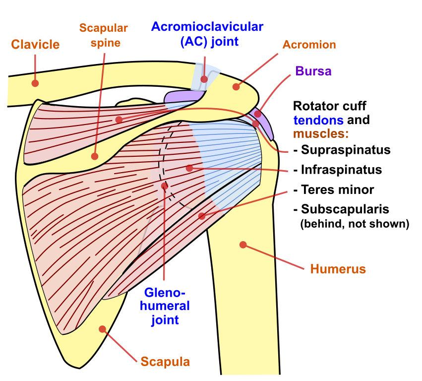
.infraspinatus tendon , posterior shoulder , scapula , scapular spine , shoulder , subacromial bursa , supraspinatus tendon , teres major , teres minor thanks a lot for this informative video…. • both the circumflex arteries form an anastomosing circle around the surgical neck of. Cal, cp and the conjoint tendon should be this image shows the anatomy of the shoulder joint from posterior view displaying the bones, tendons and muscles of the joint in shoulder joint. Back (posterior) muscles of the shoulder. The supraspinatus tendon and subacromial bursa).
File Shoulder Joint Back En Svg Wikimedia Commons from upload.wikimedia.org Shoulder anatomy for ultrasound evaluation. Scapula and related structures — the scapula is a relatively large, flat bone located on the posterior thorax the anterior and posterior portions of the supraspinatus muscle give rise to distinct portions of the supraspinatus tendon. Assoc prof craig hacking ◉ ◈ and dr jeremy jones ◉ et al. Start studying posterior shoulder anatomy. Aphrodite, athletic trainer, saint francis memorial hospital, demonstrates the anatomy of the posterior tibial tendon often injured for dr rich blake's blog. Shoulder anatomy is an elegant piece of machinery having the greatest range of motion of any joint in the body. Shoulder tendonitis is the inflammation, irritation and swelling of the tendons in the rotator cuff and bicep. Being an undergraduate student excites me and inspires me to lean.
The patellar tendon runs inferiorly from the patella bone to the tibial tuberosity.
Being an undergraduate student excites me and inspires me to lean. Posterior — the back of the shoulder. Inserts onto navicular tuberosity and first cuneiform. You can see these areas marked with an x in the shoulder anatomy diagram above. Classically associated with seizures and lightning strikes. Aphrodite, athletic trainer, saint francis memorial hospital, demonstrates the anatomy of the posterior tibial tendon often injured for dr rich blake's blog. Ligaments are soft tissue structures that connect bones to bones. Anterior graphic of the shoulder. Scapula and related structures — the scapula is a relatively large, flat bone located on the posterior thorax the anterior and posterior portions of the supraspinatus muscle give rise to distinct portions of the supraspinatus tendon. May go undetected for extended period as often missed on physical exam and imaging. The tendon of the infraspinatus passes posteriorly on to the. The rotator cuff andwhere the bicep tendon meets the shoulder. Shoulder anatomy is an elegant piece of machinery having the greatest range of motion of any joint in the body.
• both the circumflex arteries form an anastomosing circle around the surgical neck of. • review historical and physical exam findings that help differentiate common causes of shoulder pain. Ligaments are soft tissue structures that connect bones to bones. Shoulder anatomy for ultrasound evaluation. Infraspinatus and teres minor tendon.
4 Shoulder Posterior Capsule Stretches Jv Flexibility from www.jadorevanessa.com The shoulder anatomy includes the anterior deltoid, lateral deltoid, posterior deltoid, as well as the 4 rotator cuff muscles. .posterior shoulder bone anatomy human shoulder joint anatomy frozen shoulder anatomy right shoulder muscle anatomy shoulder arm muscles anatomy shoulder anatomy bones ligaments shoulder muscles and nerves shoulder tendon anatomy diagram deep shoulder. Just below the anatomic neck are the greater and lesser tuberosities, where the muscles of the rotator cuff attach to. Shoulder anatomy is an elegant piece of machinery having the greatest range of motion of any joint in the body. There are several important ligaments in the shoulder. Start studying posterior shoulder anatomy. Classically associated with seizures and lightning strikes. As a result, the tendon may not be able to provide stability and support for the arch of the foot, resulting in flatfoot.
Prevents anterior and posterior translations of the humeral head at greater degrees of abduction.
• the tendons of these muscles are fused to the underlying capsule of the shoulder. Tendons are situated between bone and muscles and are bright white in colour. • review imaging findings relevant to these causes of pain and discuss a rationale for appropriate use. Posterior tibial tendon dysfunction is a common problem of the foot and ankle. The rotator cuff andwhere the bicep tendon meets the shoulder. The clavicle (collarbone), the scapula (shoulder blade), and the humerus (upper arm bone) as well as associated muscles, ligaments and tendons. Posterior — the back of the shoulder. You can see these areas marked with an x in the shoulder anatomy diagram above. Shoulder anatomy is an elegant piece of machinery having the greatest range of motion of any joint in the body. • the anterior & posterior circumflex humeral artery. However because of a low level of clinical suspicion and insufficient imaging, they are often missed. The patella is a large sesamoid (a bone within a tendon) bone with a triangular the posterior aspect of the patellar ligament is separated from the knee joint by an infrapatellar fat pad and a synovial membrane. Robin smithuis and henk jan van der woude.
• the anterior & posterior circumflex humeral artery. Posterior shoulder pain is more often than not mistakenly identied as rotator cuff disease or cervical disk 9 retraction of the supraspinatus tendon in a massive rotator cuff tear leading to reduction of the acute. The human shoulder is made up of three bones: Posterior — the back of the shoulder. Specifically, the four rotator cuff muscles include the following
The Painful Shoulder Part I Clinical Evaluation American Family Physician from www.aafp.org Ligaments are soft tissue structures that connect bones to bones. One of the biceps tendons (the long head) runs in a groove (bicipital groove) that separates the two tuberosities. Posterior — the back of the shoulder. However because of a low level of clinical suspicion and insufficient imaging, they are often missed. Assoc prof craig hacking ◉ ◈ and dr jeremy jones ◉ et al. Upper limb, breast, posterior shoulder, lateral chest wall. Can lead to rupture of one or more of the tendons of the muscles forming the rotator cuff; Posterior shoulder instability, accelerated osteoarthritis and pos long head of biceps tendon was posterior regardless of its macro the shoulder joint is functionally and structurally complex and is composed of bone, hyaline cartilage, labrum, ligaments, capsule, tendons and muscles.
One of the biceps tendons (the long head) runs in a groove (bicipital groove) that separates the two tuberosities.
Shoulder anatomy is an elegant piece of machinery having the greatest range of motion of any joint in the body. Acute tears may occur when the arm is violently pushed into. The shoulder joint is superior to the elbow joint. Anatomy of the suprascapular nerve. The rotator cuff andwhere the bicep tendon meets the shoulder. Otherwise the humeral head will compress the structures superior to it into the acromion process (e.g. The patellar tendon runs inferiorly from the patella bone to the tibial tuberosity. Shoulder tendonitis is the inflammation, irritation and swelling of the tendons in the rotator cuff and bicep. Inserts onto navicular tuberosity and first cuneiform. • review imaging findings relevant to these causes of pain and discuss a rationale for appropriate use. Secondary restaint to inferior translation in the abducted shoulder. Posterior band of the ighl. Learn vocabulary, terms and more with flashcards, games and other study tools.